Difference between revisions of "Craniopharyngioma"
Jump to navigation
Jump to search
(redirect for now) |
(split-out) |
||
| Line 1: | Line 1: | ||
'''Craniopharyngioma''' is a benign [[neuropathology tumour]]. | |||
==General== | |||
*Develop from remains of Rathke's pouch or squamous epithelial cell rests.<ref name=pmid17425791>{{Cite journal | last1 = Garnett | first1 = MR. | last2 = Puget | first2 = S. | last3 = Grill | first3 = J. | last4 = Sainte-Rose | first4 = C. | title = Craniopharyngioma. | journal = Orphanet J Rare Dis | volume = 2 | issue = | pages = 18 | month = | year = 2007 | doi = 10.1186/1750-1172-2-18 | PMID = 17425791 }}</ref> | |||
Comes in two flavours:<ref name=pmid17425791/> | |||
*Adamantinomatous type. | |||
**Adults and children. | |||
*Squamous papillary type. | |||
**Adults individuals.<ref name=pmid6696166>{{Cite journal | last1 = Giangaspero | first1 = F. | last2 = Burger | first2 = PC. | last3 = Osborne | first3 = DR. | last4 = Stein | first4 = RB. | title = Suprasellar papillary squamous epithelioma ("papillary craniopharyngioma"). | journal = Am J Surg Pathol | volume = 8 | issue = 1 | pages = 57-64 | month = Jan | year = 1984 | doi = | PMID = 6696166 }}</ref> | |||
**Usually solid. | |||
Radiology:<ref name=pmid17425791/> | |||
*Calcified - adamantinomatous type only. | |||
*Solid & cystic. | |||
==Microscopic== | |||
===Adamantinomatous=== | |||
Features (adamantinomatous):<ref name=Ref_DCHH184>{{Ref DCHH|184}}</ref> | |||
*Well-circumscribed (or pseudoinvasive border). | |||
*Multicystic. | |||
*Small-to-medium sized cells with moderate amount of basophilic cytoplasm. | |||
*Bland nuclei (with occ. small nucleoli). | |||
*"Wet" keratin - nests of whorled keratin. | |||
*Calcifications (non-psammomatous). | |||
====Images==== | |||
<gallery> | |||
Image:Adamantinomatous_craniopharyngioma_-_very_low_mag.jpg | Adamantinomatous craniopharyngioma - very low mag. (WC/Nephron) | |||
Image:Adamantinomatous_craniopharyngioma_-_intermed_mag.jpg | Adamantinomatous craniopharyngioma - intermed. mag. (WC/Nephron) | |||
Image:Adamantinomatous_craniopharyngioma_-_very_high_mag.jpg | Adamantinomatous craniopharyngioma - very high mag. (WC/Nephron) | |||
</gallery> | |||
===Papillary=== | |||
Features (papillary):<ref name=Ref_PSNP406>{{Ref PSNP|406}}</ref> | |||
*Non-keratinized squamous epithelium (without nuclear atypia). | |||
*Fibrovascular cores (required for ''papillary''). | |||
Notes: | |||
*+/-Cilia (rare). | |||
*+/-Goblet cell-like formations (rare). | |||
====Image==== | |||
<gallery> | |||
Image:Papillary_craniopharyngioma_-_intermed_mag.jpg |Papillary craniopharyngioma - intermed. mag. (WC/Nephron) | |||
Image:Papillary_craniopharyngioma_-_very_high_mag.jpg |Papillary craniopharyngioma - very high mag. (WC/Nephron) | |||
</gallery> | |||
www: | |||
*[http://library.med.utah.edu/WebPath/jpeg4/ENDO115.jpg Craniopharyngioma (med.utah.edu)].<ref>URL: [http://library.med.utah.edu/WebPath/jpeg4/ENDO115.jpg http://library.med.utah.edu/WebPath/jpeg4/ENDO115.jpg]. Accessed on: 6 December 2010.</ref> | |||
==See also== | |||
*[[Pituitary gland]]. | |||
*[[Neuropathology tumours]]. | |||
==References== | |||
{{Reflist|2}} | |||
[[Category:Neuropathology]] | |||
Revision as of 21:09, 11 November 2013
Craniopharyngioma is a benign neuropathology tumour.
General
- Develop from remains of Rathke's pouch or squamous epithelial cell rests.[1]
Comes in two flavours:[1]
- Adamantinomatous type.
- Adults and children.
- Squamous papillary type.
- Adults individuals.[2]
- Usually solid.
Radiology:[1]
- Calcified - adamantinomatous type only.
- Solid & cystic.
Microscopic
Adamantinomatous
Features (adamantinomatous):[3]
- Well-circumscribed (or pseudoinvasive border).
- Multicystic.
- Small-to-medium sized cells with moderate amount of basophilic cytoplasm.
- Bland nuclei (with occ. small nucleoli).
- "Wet" keratin - nests of whorled keratin.
- Calcifications (non-psammomatous).
Images
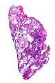
Adamantinomatous craniopharyngioma - very low mag. (WC/Nephron)
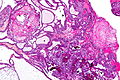
Adamantinomatous craniopharyngioma - intermed. mag. (WC/Nephron)
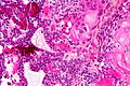
Adamantinomatous craniopharyngioma - very high mag. (WC/Nephron)


Papillary
Features (papillary):[4]
- Non-keratinized squamous epithelium (without nuclear atypia).
- Fibrovascular cores (required for papillary).
Notes:
- +/-Cilia (rare).
- +/-Goblet cell-like formations (rare).
Image
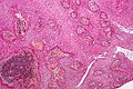
Papillary craniopharyngioma - intermed. mag. (WC/Nephron)
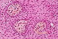
Papillary craniopharyngioma - very high mag. (WC/Nephron)


www:
See also
References
- ↑ 1.0 1.1 1.2 Garnett, MR.; Puget, S.; Grill, J.; Sainte-Rose, C. (2007). "Craniopharyngioma.". Orphanet J Rare Dis 2: 18. doi:10.1186/1750-1172-2-18. PMID 17425791.
- ↑ Giangaspero, F.; Burger, PC.; Osborne, DR.; Stein, RB. (Jan 1984). "Suprasellar papillary squamous epithelioma ("papillary craniopharyngioma").". Am J Surg Pathol 8 (1): 57-64. PMID 6696166.
- ↑ Tadrous, Paul.J. Diagnostic Criteria Handbook in Histopathology: A Surgical Pathology Vade Mecum (1st ed.). Wiley. pp. 184. ISBN 978-0470519035.
- ↑ Perry, Arie; Brat, Daniel J. (2010). Practical Surgical Neuropathology: A Diagnostic Approach: A Volume in the Pattern Recognition series (1st ed.). Churchill Livingstone. pp. 406. ISBN 978-0443069826.
- ↑ URL: http://library.med.utah.edu/WebPath/jpeg4/ENDO115.jpg. Accessed on: 6 December 2010.