Difference between revisions of "Small cell lymphomas"
(→IHC) |
|||
| Line 31: | Line 31: | ||
| mantle zone | | mantle zone | ||
| small | | small | ||
| CD5+, CD23-, CD43+, cyclin D1+<ref name=Ref_Lester95>{{Ref Lester|95}}</ref> | | CD5+, [[CD23]]-, CD43+, cyclin D1+<ref name=Ref_Lester95>{{Ref Lester|95}}</ref> | ||
| t(11;14)(q13;q32) BCL1/IGH<ref>URL: [http://atlasgeneticsoncology.org/Anomalies/t1114ID2021.html http://atlasgeneticsoncology.org/Anomalies/t1114ID2021.html]. Accessed on: 10 August 2010.</ref> (also IGH/BCL1<ref>URL: [http://www.wipo.int/patentscope/search/en/WO2010059499 http://www.wipo.int/patentscope/search/en/WO2010059499]. Accessed on: 26 May 2011.</ref>) | | t(11;14)(q13;q32) BCL1/IGH<ref>URL: [http://atlasgeneticsoncology.org/Anomalies/t1114ID2021.html http://atlasgeneticsoncology.org/Anomalies/t1114ID2021.html]. Accessed on: 10 August 2010.</ref> (also IGH/BCL1<ref>URL: [http://www.wipo.int/patentscope/search/en/WO2010059499 http://www.wipo.int/patentscope/search/en/WO2010059499]. Accessed on: 26 May 2011.</ref>) | ||
| aggressive, poor prognosis<ref name=pmid10583923/> | | aggressive, poor prognosis<ref name=pmid10583923/> | ||
| Line 56: | Line 56: | ||
| location ? | | location ? | ||
| small | | small | ||
| CD5+, CD23+, CD43+, cyclin D1- | | CD5+, [[CD23]]+, CD43+, cyclin D1- | ||
| trisomy 12; deletions of 11q, 13q, 17p<ref>{{Ref PCPBoD8|318}}</ref> | | trisomy 12; deletions of 11q, 13q, 17p<ref>{{Ref PCPBoD8|318}}</ref> | ||
| good prognosis / indolent course | | good prognosis / indolent course | ||
| Line 374: | Line 374: | ||
*CD20 +ve. | *CD20 +ve. | ||
*CD5 +ve. | *CD5 +ve. | ||
*CD23 +ve -- occasionally negative.<ref>URL: [http://path.upmc.edu/cases/case296/dx.html http://path.upmc.edu/cases/case296/dx.html]. Accessed on: 14 January 2012.</ref> | *[[CD23]] +ve -- occasionally negative.<ref>URL: [http://path.upmc.edu/cases/case296/dx.html http://path.upmc.edu/cases/case296/dx.html]. Accessed on: 14 January 2012.</ref> | ||
*CD43 +ve. | *CD43 +ve. | ||
Revision as of 20:17, 27 December 2013
The small cell lymphomas are a collection of commonly seen lymphomas that have a near-identical histomorphologic appearance.
The group includes:
- Small lymphocytic lymphoma/chronic lymphocytic leukemia.
- Follicular lymphoma.
- Mantle cell lymphoma.
- Marginal zone lymphoma (includes MALT lymphoma).
- Hairy cell leukemia.
- Immunoproliferative small intestinal disease (IPSID).[1]
Table of B-cell lymphoma
Small cell lymphomas:
| Name | Location | Size of cells | IHC | Translocations | Clinical | Other |
|---|---|---|---|---|---|---|
| Follicular lymphoma | Follicle | Small, centrocytes, centroblasts | CD10+, BCL6+[2] | t(14;18)(q32;q21) IGH/BCL2[3] | may transform into DLBCL | very common |
| Mantle cell lymphoma | mantle zone | small | CD5+, CD23-, CD43+, cyclin D1+[2] | t(11;14)(q13;q32) BCL1/IGH[4] (also IGH/BCL1[5]) | aggressive, poor prognosis[6] | DDx: Castleman disease |
| Marginal zone lymphoma (includes MALT) | marginal zone, spleen, GI tract | small | CD21+, CD11c+, CD5-, CD23-[2] | t(11;18)(q21;q21) / API2‐MALT1, t(14;18)(q32;q21) / IGH‐MALT1, t(1;14)(p22;q32) / IGH‐BCL10[7] | classical GI lymphoma | subtypes: extranodal marginal zone lymphoma (AKA MALT lymphoma), SMZL, nodal marginal zone lymphoma |
| Precursor B cell lymphoblastic lymphoma/leukemia | location ? | small | CD10+, CD5-, TdT+, CD99+[2] | t(9;22), others | good prognosis (?) | other ? |
| B cell small lymphocytic lymphoma / chronic lymphocytic leukemia |
location ? | small | CD5+, CD23+, CD43+, cyclin D1- | trisomy 12; deletions of 11q, 13q, 17p[8] | good prognosis / indolent course | other ? |
Medium and large cell lymphomas:
| Name | Location | Size of cells | IHC | Translocations | Clinical | Other |
|---|---|---|---|---|---|---|
| Burkitt's lymphoma | follicle | large cells | CD10, BCL6 | t(8;14) (q24;q32) | rapid growth | "starry sky" |
| Diffuse large B cell lymphoma | follicle (?) | large 4-5X of lymphocyte | MIB1 >40% | none/like follicular l. | poor prognosis | common among lymphomas |
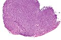
Follicular lymphoma
General
- A very common type of lymphoma.
- Expresses BCL2,[9] like many other small cell lymphomas.
Microscopic
Features (lymph node):
- Abundant abnormally-shaped lymphoid follicles - key feature - including some of the following:
- Non-polarized mantle zone (normal mantle zone is usu. thicker at capsular aspect).
- Non-polarized germinal center (normal germinal center has dark & light area).
- Loss of tingible body macrophages.
- Sinuses effaced (lost).
Note:
- The intrafollicular component of the lymph node is compressed - follicles are often described as "kissing", as they nearly touch.
- In bone marrow specimens the neoplastic cells classically have a paratrabecular arrangement,[10] i.e. the lymphoma cells are found adjacent to the bone spicules.
DDx:
- Reactive follicular hyperplasia.
- Diffuse large B-cell lymphoma - esp. for the grade 3B.
Images
Grading
- Grade 1-2: <= 22 centroblasts / HPF; where 1 HPF ~= 0.2376 mm^2 (22 mm eye piece @ 40X objective).
- Grade 3A: >22 centroblasts / HPF; where 1 HPF ~= 0.2376 mm^2 (22 mm eye piece @ 40X objective).
- Grade 3B: only centroblasts (within a nodular architecture).
Notes:
- Significant interobserver variability.[11]
- Grade 1 & Grade 2 lumped together.
- One should evaluate 10 HPFs.
- Only centroblasts without a nodular architecture is Diffuse large B cell lymphoma (DLBCL).
The usual cut points mentioned by people with HPFitis are:[12]
- Grade 1: 0-5 centroblasts / HPF.
- Grade 2: 5-15 centroblasts / HPF.
- Grade 3: >15 centroblasts / HPF.
IHC
Features:[9]
- CD10 +ve.
- BCL6 +ve.
Others:
- CD5 -ve.
- +ve in mantle cell lymphoma.
- CD23 -ve/+ve.
- +ve in CLL.
- CD43 -ve.
- +ve in mantle cell lymphoma, marginal zone lymphoma.
- CD11c -ve -- flow cytometry only.
- CD21 -ve in tumour cells; highlights follicular dendritic cells.
Image:
A panel to work-up:
- BCL2, BCL6, CD3, CD5, CD10, CD20, CD23, cyclin D1.
Molecular
- t(14;18)(q32;q21)/IGH-BCL2 in 70-95% of cases.[9]
- Should not be confused with t(14;18)(q32;q21)/IGH-MALT1 seen in MALT lymphomas.[7]
Sign out
RETROPERITONEAL MASS, RIGHT, CORE BIOPSIES:
- NON-HODGKIN B-CELL LYMPHOMA, FAVOUR FOLLICULAR LYMPHOMA.

COMMENT:
Morphology:
-Small cells: size ~ mature lymphocytes, quantity - many, angular and round.
-Large cells (intermixed with small cells): size ~1.5-2x mature lymphocyte,
small nucleoli, moderate quantity of grey/basophilic cytoplasm, moderate
nuclear pleomorphism.
-Architecture: no gland formation, discohesive, no follicles apparent, no sheets
of large cells.
-Mitoses are uncommon.

Immunohistochemical stains (tumour cells):
Positive: CD45 (strong, membranous/cytoplasmic), CD20 (strong, membranous/cytoplasmic),
BCL-2 (strong, membranous/cytoplasmic), CD10 (strong, membranous), BCL-6 (moderate,
patchy, nuclear).

Negative: pankeratin, CD3, CD5, CD30, CD21 (follicular dendritic cells not apparent),
CD23 (scattered, rare).

Ki-67: highlights the large cells, primarily -- 5-35% of cells within the core.

The findings favour a follicular lymphoma, based on the cellular morphology and
immunostains; however, they are limited by the type of tissue sampling (core biopsy).
Clinical correlation is suggested.
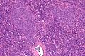
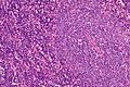
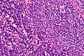
Mantle cell lymphoma
- Abbreviated MCL.
General
- Relatively aggressive - guarded prognosis.[6]
Microscopic
Features:[13]
- Small monomorphic (uniform size, shape and staining) lymphoid population.
- Abundant mitoses.
- Scattered epithelioid histiocytes (should not be confused with tingible-body macrophages).
- Sclerosed blood vessels.
DDx:
- Other small cell lymphomas, esp. marginal zone lymphoma.
- Burkitt's lymphoma.
Images:
IHC
Others:
- CD23 -ve.
- Positive in CLL.
Molecular
- t(11;14)(q13;q32) / IGH-CCND1.[16]
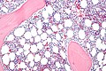
Marginal zone lymphoma
General
- Abbreviated as MZL.
- Arise in the context chronic infections, e.g. Sjögren disease (salivary gland), Hashimoto thyroiditis (thyroid gland), Helicobacter pylori gastritis (stomach).[17]
Classification
- Comes in three different flavours:
- Extranodal marginal zone lymphoma.
- If in mucosa-associated lymphoid tissue known as a MALT lymphoma, AKA MALToma.
- Splenic marginal zone lymphoma (SMZL).
- Nodal marginal zone lymphoma (NMZL).
- Extranodal marginal zone lymphoma.
Microscopic
Features:
- Small (lymphoid) cells that may be plasma cell-like (plasmacytoid):[18]
- +/-Clockface nucleus.
- +/-Eccentric nucleus.
- +/-"Lymphoepithelial lesion" - gastric crypts invaded by a monomorphous population of lymphocytes.[19]
- Features:
- Cluster of lymphocytes - three cells or more - key feature.
- Single lymphocytes don't count.
- Clearing around the lymphocyte cluster.
- Cluster of lymphocytes - three cells or more - key feature.
- Not specific for MALT lymphoma, i.e. may be seen in other types of lymphoma.[20]
- Features:
Images

Lymphoepithelial lesion - very high mag. (WC)
Lymphoepithelial lesion - intermed. mag. (WC)


www:
IHC
Features:[21]
- CD20 +ve.
- BCL2 +ve.
- CD21 +ve.
- CD11c +ve (flow cytometry or laser scanning cytometry - only; not available for paraffin).
- CD43 +ve/-ve.
- Typically positive in mantle cell lymphoma.
Others:
- CD5 -ve.
- CD10 -ve.
- CD23 -ve.
Molecular
There are several associated with MALT lymphoma:[7]
- t(11;18)(q21;q21) / API2‐MALT1[22] - most common translocation in MALT lymphoma.[23]
- t(14;18)(q32;q21) / IGH‐MALT1.
- Should not be confused with t(14;18) seen in follicular lymphoma between IGH-BCL2.[9]
- t(1;14)(p22;q32) / IGH‐BCL10.
The MALT1 associated translocations can be assessed with an ISH break apart probe for MALT1.
Hairy cell leukemia
- Abbreviated HCL.
General
- Name comes from appearance on blood smear - cell hairy.
- Do to the biology, dry taps are common.[24]
Clinical:[25]
- Pancytopenia.
- Splenic enlargement.
- No lymphadenopathy.
- Good prognosis (with treatment), though (likely) not curable.
Gross
Features:[17]
- Huge beefy red spleen.
- Red as white pulp obliterated.
Microscopic
Features:[26]
- Small cells (10-20 micrometers) with "Fried egg"-like appearance:
- Well-demarcated fuzzy cell borders,
- Clear/whispy cytoplasm and,
- Central round nucleus.
- Peri-nuclear clearing ("water-clear rim"[27]) -- key feature.
DDx:
Images
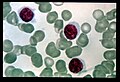
HCL - blood film. (WC)
HCL - high mag. (WC)

HCL - very high mag. (WC)


www:
- HCL - bone marrow (nlm.nih.gov) from Holland-Frei Cancer Medicine (nlm.nih.gov).
- HCL - several images (upmc.edu).
- HCL - another case with several images (upmc.edu).
- HCL in the spleen (webpathology.com).
IHC
Features:[28]
- CD20 +ve, CD25 +ve, CD103 +ve.
- CD5 -ve.
Flow cytometry:
- CD19 +ve, CD11c +ve, FMC7 +ve.
B cell small lymphocytic lymphoma/chronic lymphocytic leukemia
- Abbreviated CLL and SLL.
General
- Very common.
- Good prognosis.
Richter's transformation
- CLL/SLL may under go a Richter's transformation into a high-grade non-Hodgkin's lymphoma (NHL), e.g. DLBCL:[29]
- Incidence of transformation <5%.
- Prognosis < 1 year.
Microscopic
Features in a lymph node:[30]
- Mixed population of lymphoid cells with "proliferation centers" - key feature:
- Larger cells (~ 1.5x the size of resting lymphocyte ~ 12-15 micrometers):
- Nucleoli.
- Form (nodular) collections.
- Small dark cells (~ size of resting lymphocyte ~ 8-10 micrometers):
- Predominant population.
- Lack nucleolus.
- Larger cells (~ 1.5x the size of resting lymphocyte ~ 12-15 micrometers):
Images
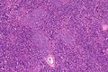
CLL - low mag. (WC)
CLL - intermed. mag. (WC)
CLL - high mag. (WC)
CLL - very high mag. (WC)


www:
- CLL in the liver (pathologyatlas.ro).
- SLL in the parotid - several images (upmc.edu).
- CLL with Richter transformation - several images (upmc.edu).
IHC
Others:
- Cyclin D1 -ve.
Molecular
- Lacks t(11;14) seen in mantle cell lymphoma.
Precursor B-cell lymphoblastic lymphoma/leukemia
General
- Good prognosis.
- Paediatric - usu. <6 years old.
Microscopic
Features:[32]
- High mitotic rate.
- "Starry sky" pattern.
- Small nucleoli.
IHC
Features:[2]
- CD10 +ve, TdT +ve, CD99 +ve.
- CD5 -ve.
Molecular
Subclassification based on molecular abnormalities (translocations, rearrangements):[33]
- t(9;22) / BCR-ABL.
- t(1;19) / E2A-PBX1.
- t(12;21) / ETV-CBFalpha.
- MLL rearrangement.
Precursor T-cell lymphoblastic lymphoma/leukemia
General
- Prognosis poor. (???)
Microscopic
Features:
- Small lymphoid cells. (???)
IHC
Features:[34]
- TdT +ve, CD34 +ve, CD99 +ve, CD1a +ve/-ve.
- TIA1 -ve.
See also
References
- ↑ Al-Saleem T, Al-Mondhiry H (March 2005). "Immunoproliferative small intestinal disease (IPSID): a model for mature B-cell neoplasms". Blood 105 (6): 2274–80. doi:10.1182/blood-2004-07-2755. PMID 15542584. http://bloodjournal.hematologylibrary.org/cgi/content/long/105/6/2274.>
- ↑ 2.0 2.1 2.2 2.3 2.4 Lester, Susan Carole (2005). Manual of Surgical Pathology (2nd ed.). Saunders. pp. 95. ISBN 978-0443066450.
- ↑ Yanai, S.; Nakamura, S.; Takeshita, M.; Fujita, K.; Hirahashi, M.; Kawasaki, K.; Kurahara, K.; Sakai, Y. et al. (Dec 2010). "Translocation t(14;18)/IGH-BCL2 in gastrointestinal follicular lymphoma: correlation with clinicopathologic features in 48 patients.". Cancer. doi:10.1002/cncr.25811. PMID 21192062.
- ↑ URL: http://atlasgeneticsoncology.org/Anomalies/t1114ID2021.html. Accessed on: 10 August 2010.
- ↑ URL: http://www.wipo.int/patentscope/search/en/WO2010059499. Accessed on: 26 May 2011.
- ↑ 6.0 6.1 Hankin, RC.; Hunter, SV. (Dec 1999). "Mantle cell lymphoma.". Arch Pathol Lab Med 123 (12): 1182-8. doi:10.1043/0003-9985(1999)1231182:MCL2.0.CO;2. PMID 10583923.
- ↑ 7.0 7.1 7.2 Bacon CM, Du MQ, Dogan A (April 2007). "Mucosa-associated lymphoid tissue (MALT) lymphoma: a practical guide for pathologists". J. Clin. Pathol. 60 (4): 361–72. doi:10.1136/jcp.2005.031146. PMC 2001121. PMID 16950858. https://www.ncbi.nlm.nih.gov/pmc/articles/PMC2001121/.
- ↑ Mitchell, Richard; Kumar, Vinay; Fausto, Nelson; Abbas, Abul K.; Aster, Jon (2011). Pocket Companion to Robbins & Cotran Pathologic Basis of Disease (8th ed.). Elsevier Saunders. pp. 318. ISBN 978-1416054542.
- ↑ 9.0 9.1 9.2 9.3 Vitolo U, Ferreri AJ, Montoto S (June 2008). "Follicular lymphomas". Crit. Rev. Oncol. Hematol. 66 (3): 248–61. doi:10.1016/j.critrevonc.2008.01.014. PMID 18359244.
- ↑ Iancu, D.; Hao, S.; Lin, P.; Anderson, SK.; Jorgensen, JL.; McLaughlin, P.; Medeiros, LJ. (Feb 2007). "Follicular lymphoma in staging bone marrow specimens: correlation of histologic findings with the results of flow cytometry immunophenotypic analysis.". Arch Pathol Lab Med 131 (2): 282-7. doi:10.1043/1543-2165(2007)131[282:FLISBM]2.0.CO;2. PMID 17284114.
- ↑ DG. 17 August 2010.
- ↑ Mills, Stacey E; Carter, Darryl; Greenson, Joel K; Oberman, Harold A; Reuter, Victor E (2004). Sternberg's Diagnostic Surgical Pathology (4th ed.). Lippincott Williams & Wilkins. pp. 813. ISBN 978-0781740517.
- ↑ DG. 17 August 2010.
- ↑ URL: http://path.upmc.edu/cases/case308/dx.html. Accessed on: 14 January 2012.
- ↑ Online 'Mendelian Inheritance in Man' (OMIM) 168461
- ↑ URL: http://atlasgeneticsoncology.org/Anomalies/t1114ID2021.html. Accessed on: 10 August 2010.
- ↑ 17.0 17.1 Mitchell, Richard; Kumar, Vinay; Fausto, Nelson; Abbas, Abul K.; Aster, Jon (2011). Pocket Companion to Robbins & Cotran Pathologic Basis of Disease (8th ed.). Elsevier Saunders. pp. 326. ISBN 978-1416054542.
- ↑ URL: http://surgpathcriteria.stanford.edu/bcell/marginalnodal/printable.html. Accessed on: 6 March 2012.
- ↑ Papadaki, L.; Wotherspoon, AC.; Isaacson, PG. (Nov 1992). "The lymphoepithelial lesion of gastric low-grade B-cell lymphoma of mucosa-associated lymphoid tissue (MALT): an ultrastructural study.". Histopathology 21 (5): 415-21. PMID 1452124.
- ↑ DB. 6 August 2010.
- ↑ Lester, Susan Carole (2005). Manual of Surgical Pathology (2nd ed.). Saunders. pp. 95. ISBN 978-0443066450.
- ↑ Mitchell, Richard; Kumar, Vinay; Fausto, Nelson; Abbas, Abul K.; Aster, Jon (2011). Pocket Companion to Robbins & Cotran Pathologic Basis of Disease (8th ed.). Elsevier Saunders. pp. 170. ISBN 978-1416054542.
- ↑ Streubel, B.; Lamprecht, A.; Dierlamm, J.; Cerroni, L.; Stolte, M.; Ott, G.; Raderer, M.; Chott, A. (Mar 2003). "T(14;18)(q32;q21) involving IGH and MALT1 is a frequent chromosomal aberration in MALT lymphoma.". Blood 101 (6): 2335-9. doi:10.1182/blood-2002-09-2963. PMID 12406890.
- ↑ Galani, KS.; Subramanian, PG.; Gadage, VS.; Rahman, K.; Ashok Kumar, MS.; Shinde, S.; Mahadik, S.; Ansari, R. et al. "Clinico-pathological profile of Hairy cell leukemia: critical insights gained at a tertiary care cancer hospital.". Indian J Pathol Microbiol 55 (1): 61-5. doi:10.4103/0377-4929.94858. PMID 22499303.
- ↑ URL: http://www.ncbi.nlm.nih.gov/bookshelf/br.fcgi?book=cmed&part=A34022. Accessed on: 20 August 2010.
- ↑ URL: http://emedicine.medscape.com/article/200580-diagnosis. Accessed on: 18 August 2010.
- ↑ URL: http://www.ncbi.nlm.nih.gov/bookshelf/br.fcgi?book=cmed&part=A34022. Accessed on: 20 August 2010.
- ↑ URL: http://www.ncbi.nlm.nih.gov/bookshelf/br.fcgi?book=cmed&part=A34022&rendertype=table&id=A34029. Accessed on: 20 August 2010.
- ↑ Tsimberidou AM, Keating MJ (April 2006). "Richter's transformation in chronic lymphocytic leukemia". Semin. Oncol. 33 (2): 250–6. doi:10.1053/j.seminoncol.2006.01.016. PMID 16616072.
- ↑ DG. 17 August 2010.
- ↑ URL: http://path.upmc.edu/cases/case296/dx.html. Accessed on: 14 January 2012.
- ↑ DG. 17 August 2010.
- ↑ Randolph TR (2004). "Advances in acute lymphoblastic leukemia". Clin Lab Sci 17 (4): 235–45. PMID 15559730. http://findarticles.com/p/articles/mi_qa3890/is_200410/ai_n9429273/pg_2.
- ↑ Lester, Susan Carole (2005). Manual of Surgical Pathology (2nd ed.). Saunders. pp. 97. ISBN 978-0443066450.