Difference between revisions of "CNS cytopathology"
m (→Basic approach) |
Jensflorian (talk | contribs) |
||
| (11 intermediate revisions by 3 users not shown) | |||
| Line 2: | Line 2: | ||
An introduction to cytopathology is in the ''[[cytopathology]]'' article. Cerebrospinal (CSF) specimens are dealt with in a separate article called ''[[CSF cytopathology]]''. | An introduction to cytopathology is in the ''[[cytopathology]]'' article. Cerebrospinal (CSF) specimens are dealt with in a separate article called ''[[CSF cytopathology]]''. | ||
==Technique== | |||
Smears (really squash preps) are common in neuropathology. Here are some tips for getting a good smear: | |||
# Sampling is key -- choose 3-4 small pieces of tissue from different areas of the tissue (if there are different colours, get some of each). | |||
# Keep pieces small (easier to smear). | |||
# Avoid air drying (place into formal alcohol immediately upon smearing). | |||
==Basic approach== | ==Basic approach== | ||
| Line 11: | Line 16: | ||
{{familytree | C01 | | | | C02 | | C03 | | | | C04 | |C01=Glial|C02=Non-glial|C03=Infectious|C04=Non-infectious }} | {{familytree | C01 | | | | C02 | | C03 | | | | C04 | |C01=Glial|C02=Non-glial|C03=Infectious|C04=Non-infectious }} | ||
{{familytree/end}} | {{familytree/end}} | ||
{| class="wikitable" | |||
| | |||
| Glial | |||
| Non-glial | |||
|- | |||
| Stranding <br>(cytoplasmic) | |||
| thin - cannot be seen at low <br>power (2.5x obj.), true cytoplasmic <br>processes | |||
| thick - can be seen at low <br>power (2.5x obj.), artifact of smearing | |||
|- | |||
| Edge of cluster | |||
| smooth/non-distinct | |||
| sharp | |||
|} | |||
Glial vs non-glial: | Glial vs non-glial: | ||
*Glial has | *Glial has cytoplasmic processes/cytoplasmic strands (stringy processes) ~ 1 micrometer thick. | ||
**They cannot be seen well at low power. | |||
**Cotton candy-like appearance. | |||
**Images: | **Images: | ||
*** [http://www.msdlatinamerica.com/ebooks/DiagnosticNeuropathologySmears/files/cf76aad0d9dab70c99b93186ca8b3ad4.gif Stringy processes - glial tumour (msdlatinamerica.com)].<ref name=latinam>URL: [http://www.msdlatinamerica.com/ebooks/DiagnosticNeuropathologySmears/sid117213.html http://www.msdlatinamerica.com/ebooks/DiagnosticNeuropathologySmears/sid117213.html]. Accessed on: 2 November 2010.</ref> | *** [http://www.msdlatinamerica.com/ebooks/DiagnosticNeuropathologySmears/files/cf76aad0d9dab70c99b93186ca8b3ad4.gif Stringy processes - glial tumour (msdlatinamerica.com)].<ref name=latinam>URL: [http://www.msdlatinamerica.com/ebooks/DiagnosticNeuropathologySmears/sid117213.html http://www.msdlatinamerica.com/ebooks/DiagnosticNeuropathologySmears/sid117213.html]. Accessed on: 2 November 2010.</ref> | ||
*** [http://www.msdlatinamerica.com/ebooks/DiagnosticNeuropathologySmears/files/59f74df1106573dcdea73be7febff2aa.gif Glial tumour mimicing a carcinoma (msdlatinamerica.com)].<ref name=latinam>URL: [http://www.msdlatinamerica.com/ebooks/DiagnosticNeuropathologySmears/sid117213.html http://www.msdlatinamerica.com/ebooks/DiagnosticNeuropathologySmears/sid117213.html]. Accessed on: 2 November 2010.</ref> | *** [http://www.msdlatinamerica.com/ebooks/DiagnosticNeuropathologySmears/files/59f74df1106573dcdea73be7febff2aa.gif Glial tumour mimicing a carcinoma (msdlatinamerica.com)].<ref name=latinam>URL: [http://www.msdlatinamerica.com/ebooks/DiagnosticNeuropathologySmears/sid117213.html http://www.msdlatinamerica.com/ebooks/DiagnosticNeuropathologySmears/sid117213.html]. Accessed on: 2 November 2010.</ref> | ||
Gliosis vs. neoplasm: | |||
*Gliosis - uniform, pink | |||
*Astrocytoma - irregular, coarse clumps of pink fibrillary material | |||
High grade vs. low-grade: | |||
*Markers of high grade glioma: | |||
# Mitoses (can see these in smear) | |||
# Necrosis (can also see in smears) | |||
Notes: | Notes: | ||
| Line 25: | Line 55: | ||
***Crushed/elongated nuclei are present in artifactual processes. | ***Crushed/elongated nuclei are present in artifactual processes. | ||
<!-- *** something i forgot --> | <!-- *** something i forgot --> | ||
===Meningioma=== | |||
Most meningiomas smear rather well, the only exception being ones that are densely fibrous. | |||
*Key features of meningioma smears: | |||
# Single cells or small groups of epithelioid cells with distinct cytoplasmic 'flags' (cytoplasm usually abundant) | |||
# Round nuclei with vesicular chromatin and unapparent or small nucleoli | |||
# Visible actin striations / stress filaments in cells (seen as pink straight lines) | |||
<gallery> | |||
File:Meningioma cytologie.jpg | HE smear of a meningioma displaying typical whorl formations (WC/jensflorian) | |||
</gallery> | |||
===Metastatic carcinoma=== | ===Metastatic carcinoma=== | ||
Typically has a 'cannonball' appearance -- with small, highly cohesive clusters of epithelioid cells. | |||
{{Main|Metastasis}} | |||
*[http://moon.ouhsc.edu/kfung/JTY1/NeuroTest/_derived/Q92-Ans.htm_txt_SampleQ92.gif Squamous cell carcinoma (ouhsc.edu)].<ref>URL: [http://moon.ouhsc.edu/kfung/JTY1/NeuroTest/Q92-Ans.htm http://moon.ouhsc.edu/kfung/JTY1/NeuroTest/Q92-Ans.htm]. Accessed on: 3 November 2010.</ref> | *[http://moon.ouhsc.edu/kfung/JTY1/NeuroTest/_derived/Q92-Ans.htm_txt_SampleQ92.gif Squamous cell carcinoma (ouhsc.edu)].<ref>URL: [http://moon.ouhsc.edu/kfung/JTY1/NeuroTest/Q92-Ans.htm http://moon.ouhsc.edu/kfung/JTY1/NeuroTest/Q92-Ans.htm]. Accessed on: 3 November 2010.</ref> | ||
*[http://moon.ouhsc.edu/kfung/JTY1/NeuroTest/Images/SampleQ50.gif | *[http://moon.ouhsc.edu/kfung/JTY1/NeuroTest/Images/SampleQ50.gif Adenocarcinoma (ouhsc.edu)]. | ||
*[http://moon.ouhsc.edu/kfung/JTY1/NeuroTest/Images/SampleQ59.gif Small cell carcinoma (ouhsc.edu)]. | *[http://moon.ouhsc.edu/kfung/JTY1/NeuroTest/Images/SampleQ59.gif Small cell carcinoma (ouhsc.edu)]. | ||
===Metastatic melanoma=== | |||
*Occasionally large bi- or multinucleated cells | |||
*Discohesive cells | |||
*Large nucleoli | |||
*Pigmented tumor cells | |||
*Accompanied by tumor infiltrating lymphocytes | |||
<gallery> | |||
File:Melanoma_CNS_met_smear_HE.jpg | Melanoma smear prep (H&E) | |||
File:Melanoma - cytology field stain.jpg | |||
</gallery> | |||
===Metastatic lymphoma=== | |||
*Small round and blue cells | |||
===Glial tumors=== | |||
* Elongated cell processes | |||
* Cohesive growth | |||
* Often attached to vasculature | |||
<gallery> | |||
File:Pilocytic astrocytoma - smear - very high mag.jpg | Pilocytic astrocytoma smear preparation (H&E) | |||
</gallery> | |||
==Things that don't smear well== | ==Things that don't smear well== | ||
Cohesive tumours: | Cohesive tumours: | ||
* | *[[Neurofibroma]]. | ||
* | *[[Schwannoma]].<ref name=Ref_TPoSP|252>{{Ref TPoSP|252}}</ref> | ||
* | *Subependymoma | ||
*Abscess (because of fibrous capsule) | |||
*Metastasis.<ref name=Ref_TPoSP|252>{{Ref TPoSP|252}}</ref> | *Metastasis.<ref name=Ref_TPoSP|252>{{Ref TPoSP|252}}</ref> | ||
===Things that smear well=== | ===Things that smear well=== | ||
Dyscohesive tumours:<ref name=Ref_TPoSP|252>{{Ref TPoSP|252}}</ref> | Dyscohesive tumours: | ||
* | *[[Lymphoma]].<ref name=Ref_TPoSP|252>{{Ref TPoSP|252}}</ref> | ||
* | *[[Pituitary adenoma]].<ref name=Ref_TPoSP|252>{{Ref TPoSP|252}}</ref> | ||
* | *Oligodendroglioma.<ref name=Ref_TPoSP|252>{{Ref TPoSP|252}}</ref> | ||
*[[Astrocytoma]]. | |||
*Normal brain. | |||
==See also== | ==See also== | ||
Latest revision as of 14:59, 21 December 2016
CNS cytopathology is a subset of neuropathology and cytopathology.
An introduction to cytopathology is in the cytopathology article. Cerebrospinal (CSF) specimens are dealt with in a separate article called CSF cytopathology.
Technique
Smears (really squash preps) are common in neuropathology. Here are some tips for getting a good smear:
- Sampling is key -- choose 3-4 small pieces of tissue from different areas of the tissue (if there are different colours, get some of each).
- Keep pieces small (easier to smear).
- Avoid air drying (place into formal alcohol immediately upon smearing).
Basic approach
| CNS cytology | |||||||||||||||||||||||||||||||||||||||||
| Tumour | Non-tumour | ||||||||||||||||||||||||||||||||||||||||
| Glial | Non-glial | Infectious | Non-infectious | ||||||||||||||||||||||||||||||||||||||
| Glial | Non-glial | |
| Stranding (cytoplasmic) |
thin - cannot be seen at low power (2.5x obj.), true cytoplasmic processes |
thick - can be seen at low power (2.5x obj.), artifact of smearing |
| Edge of cluster | smooth/non-distinct | sharp |
Glial vs non-glial:
- Glial has cytoplasmic processes/cytoplasmic strands (stringy processes) ~ 1 micrometer thick.
- They cannot be seen well at low power.
- Cotton candy-like appearance.
- Images:
Gliosis vs. neoplasm:
- Gliosis - uniform, pink
- Astrocytoma - irregular, coarse clumps of pink fibrillary material
High grade vs. low-grade:
- Markers of high grade glioma:
- Mitoses (can see these in smear)
- Necrosis (can also see in smears)
Notes:
- Crush artifact (in smear preparation) can mimic glial processes.
- Crush artifact vs. real glial processes:
- No glial processes run perpendicular to the direction of smear.
- Glial processes may branch.
- Crushed/elongated nuclei are present in artifactual processes.
- Crush artifact vs. real glial processes:
Meningioma
Most meningiomas smear rather well, the only exception being ones that are densely fibrous.
- Key features of meningioma smears:
- Single cells or small groups of epithelioid cells with distinct cytoplasmic 'flags' (cytoplasm usually abundant)
- Round nuclei with vesicular chromatin and unapparent or small nucleoli
- Visible actin striations / stress filaments in cells (seen as pink straight lines)
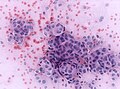
HE smear of a meningioma displaying typical whorl formations (WC/jensflorian)

Metastatic carcinoma
Typically has a 'cannonball' appearance -- with small, highly cohesive clusters of epithelioid cells.
- Squamous cell carcinoma (ouhsc.edu).[2]
- Adenocarcinoma (ouhsc.edu).
- Small cell carcinoma (ouhsc.edu).
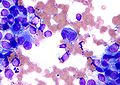
Metastatic melanoma
- Occasionally large bi- or multinucleated cells
- Discohesive cells
- Large nucleoli
- Pigmented tumor cells
- Accompanied by tumor infiltrating lymphocytes
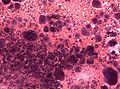
Melanoma smear prep (H&E)


Metastatic lymphoma
- Small round and blue cells
Glial tumors
- Elongated cell processes
- Cohesive growth
- Often attached to vasculature
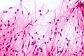
Pilocytic astrocytoma smear preparation (H&E)

Things that don't smear well
Cohesive tumours:
- Neurofibroma.
- Schwannoma.[3]
- Subependymoma
- Abscess (because of fibrous capsule)
- Metastasis.[3]
Things that smear well
Dyscohesive tumours:
- Lymphoma.[3]
- Pituitary adenoma.[3]
- Oligodendroglioma.[3]
- Astrocytoma.
- Normal brain.
See also
References
- ↑ 1.0 1.1 URL: http://www.msdlatinamerica.com/ebooks/DiagnosticNeuropathologySmears/sid117213.html. Accessed on: 2 November 2010.
- ↑ URL: http://moon.ouhsc.edu/kfung/JTY1/NeuroTest/Q92-Ans.htm. Accessed on: 3 November 2010.
- ↑ 3.0 3.1 3.2 3.3 3.4 Weedman Molavi, Diana (2008). The Practice of Surgical Pathology: A Beginner's Guide to the Diagnostic Process (1st ed.). Springer. pp. 252. ISBN 978-0387744858.