Difference between revisions of "Embryonal tumour with multilayered rosettes"
Jump to navigation
Jump to search
Jensflorian (talk | contribs) m (see also) |
Jensflorian (talk | contribs) (→Microscopy: +images) |
||
| Line 34: | Line 34: | ||
* Papillar and tubular growth (primitive neural tubes). | * Papillar and tubular growth (primitive neural tubes). | ||
**PAS-positive membranes. | **PAS-positive membranes. | ||
* Glial maturation after treatment (rare). | * Glial/neuronal maturation after treatment (rare). | ||
<gallery> | |||
File:Medulloepithelioma Histology.jpg | ETMR, medulloepithelioma type. | |||
File:Ependymoblastoma.jpg | ETMR, ependymoblastoma type. | |||
File:Ependymoblastoma ETMRjpg.jpg | ETMR, poorly undifferentiated cells. | |||
File:Neuroblasts ependymoblastoma.jpg | ETMR, ganglion cells. | |||
==IHC== | ==IHC== | ||
Revision as of 12:04, 4 October 2017
Embryonal tumour with multilayered rosettes, abbreviated ETMR, is a very rare neuropathology embryonal tumour with aggressive behaviour.
General
- Extremely rare.
- ETMR historically had been termed CNS PNET.
- The WHO2016 CNS classification contains two groups:
- Embryonal tumour with multilayered rosettes, C19MC-altered.
- Embryonal tumour with multilayered rosettes, NOS.
Note: ETMR is an umbrella term for tumors formerly known as:[1]
- Embryonal tumour with abundant neuropil and true rosettes [2]
- Ependymoblastoma [3]
- Medulloepithelioma[4]
Clinical presentation
- Usu. age <4 years.
- 70% supratentorial, 30% infratentorial.
- Raised intracranial pressure.
Imaging
- Usu. enhancing.
- Rarely cysts, calcifications.
- Widespread infiltration.
Microscopy
- Rosettes (often multilayered).
- Small cells.
- Fibrillar zones (neuropil-like areas).
- Neoplastic ganglion cells.
- Papillar and tubular growth (primitive neural tubes).
- PAS-positive membranes.
- Glial/neuronal maturation after treatment (rare).
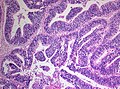
ETMR, medulloepithelioma type.
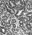
ETMR, ependymoblastoma type.
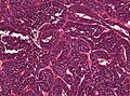
ETMR, poorly undifferentiated cells.
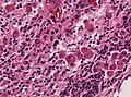
ETMR, ganglion cells.
- ==IHC==
- *LIN28+ve.
- *CD99: focally +ve.
- *Synaptophysin: Neuropil-like areas +ve.
- *GFAP: usu -ve.
- *INI1 +ve.
- *Mib1: 20-80%.


See also
References
- ↑ Korshunov, A.; Sturm, D.; Ryzhova, M.; Hovestadt, V.; Gessi, M.; Jones, DT.; Remke, M.; Northcott, P. et al. (Aug 2014). "Embryonal tumor with abundant neuropil and true rosettes (ETANTR), ependymoblastoma, and medulloepithelioma share molecular similarity and comprise a single clinicopathological entity.". Acta Neuropathol 128 (2): 279-89. doi:10.1007/s00401-013-1228-0. PMID 24337497.
- ↑ Ceccom, J.; Bourdeaut, F.; Loukh, N.; Rigau, V.; Milin, S.; Takin, R.; Richer, W.; Uro-Coste, E. et al. "Embryonal tumor with multilayered rosettes: diagnostic tools update and review of the literature.". Clin Neuropathol 33 (1): 15-22. doi:10.5414/NP300636. PMID 23863344.
- ↑ Judkins, AR.; Ellison, DW. (Jan 2010). "Ependymoblastoma: dear, damned, distracting diagnosis, farewell!*.". Brain Pathol 20 (1): 133-9. doi:10.1111/j.1750-3639.2008.00253.x. PMID 19120373.
- ↑ Korshunov, A.; Jakobiec, FA.; Eberhart, CG.; Hovestadt, V.; Capper, D.; Jones, DT.; Sturm, D.; Stagner, AM. et al. (Dec 2015). "Comparative integrated molecular analysis of intraocular medulloepitheliomas and central nervous system embryonal tumors with multilayered rosettes confirms that they are distinct nosologic entities.". Neuropathology 35 (6): 538-44. doi:10.1111/neup.12227. PMID 26183384.