Suture material
Suture material is occasionally seen under the microscope. It is usually easy to identified and typically polarizes.
General
- Suture are often used to orient a specimen[1] and/or mark the true surgical margin.[2]
Microscopic
Features:
- Glassy appearance - sharply circumscribed.
- +/-Tearing surrounding tissue.
- +/-Foreign body-type granulomas with multinucleated giant cells.
- Seen only if the suture has been in place for at least several days.
Images
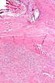
Suture material adjacent to a SFT.
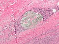
Suture material.


See also
References
- ↑ Volleamere, AJ.; Kirwan, CC. (Mar 2013). "National survey of breast cancer specimen orientation marking systems.". Eur J Surg Oncol 39 (3): 255-9. doi:10.1016/j.ejso.2012.12.008. PMID 23287819.
- ↑ Seitz, SE.; Foley, GL.; Marretta, SM. (Jun 1995). "Evaluation of marking materials for cutaneous surgical margins.". Am J Vet Res 56 (6): 826-33. PMID 7544556.