Ependymoma
Revision as of 15:04, 15 April 2015 by Jensflorian (talk | contribs) (minor update. I suggest moving myxopapillary ependymoma into a separate page)
Ependymoma is a neuropathology tumour.
General
- Called the forgotten glial tumour.
Epidemiology:[1]
- Usual site:
- Adults: usu. spinal cord.
- Children: usu. posterior fossa.
- May be assoc. with neurofibromatosis 2.
There are four main ependymal tumors:
- Ependymoma (not otherwise specified).
- Other flavours:[2]
- Cellular ependymoma.
- Papillary ependymoma.
- Clear cell ependymoma.
- Tanycytic ependymoma.
- Other flavours:[2]
- Anaplastic ependymoma.
- Myxopapillary ependymoma.
- Classically at filum terminale.
- Subependymoma
- Typically seen in IVth ventricle
Microscopic
Classic ependymoma
Features:
- Cells have a "tadpole-like" morphology.
- May also be described as ice cream cone-shaped.[3]
- Rosettes = circular nuclear free zones/cells arranged in a pseudoglandular fashion; comes in two flavours in ependymoma:
- Perivascular pseudorosettes = (tumour) cells arranged around a blood vessel; nuclei of cells distant from the blood vessel, i.e. rim of cytoplasm (from tumour cells) surround blood vessel (nucleus-free zone); more common than ependymal rosette... but less specific.
- Ependymal rosette (AKA true ependymal rosette) = rosette has an empty space at the centre - key feature.
- Nuclear features monotonous, i.e. "boring".[4]
- There is little variation in size, shape and staining.
DDx (classic ependymoma):
- Subependymoma.
- Glioblastoma (GBM).
- Invasive border = GBM; circumscribed border of lesion = ependymoma.
Images
www:
- Ependymoma (flickr.com).
- Ependymoma - ependymal rosettes (ajnr.org).
- Anaplastic ependymoma - case 1 (upmc.edu).
- Anaplastic ependymoma - case 2 (upmc.edu).
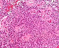
Ependymoma - intermed. mag. (WC)
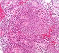
Ependymoma - low mag. (WC)


Myxopapillary ependymoma
Features:
- Perivascular pseudorosettes:
- Myxoid material surround blood vessels.
- Myxoid material surrounded by tumour cells.
- Myxoid material surround blood vessels.
Images:
- Myxopapillary ependymoma - high mag. (WC).
- Myxopapillary ependymoma (bmj.com) - part of careers.bmj.com article on paediatric pathology.
- Myxopapillary ependymoma - cytology (WC).
- Myxopapillary ependymoma - several images (upmc.edu).
Grading
Easy:
- Subependymoma = WHO grade I.
- Myxopapillary ependymoma = WHO grade I.
Not-so-easy:
- Classic ependymoma = WHO grade II.
- Anaplastic ependymoma = WHO grade III.
Grade II vs. Grade III:
- Cellular density.
- Mitoses.
- Necrosis.
- Microvascular proliferation.
Notes:
- Many tumours fall between grade II and grade III. These are called "indeterminate" by many.
IHC
- Reticulin.
- GFAP.
- MIB1.
See also
References
- ↑ Kumar, Vinay; Abbas, Abul K.; Fausto, Nelson; Aster, Jon (2009). Robbins and Cotran pathologic basis of disease (8th ed.). Elsevier Saunders. pp. 1334. ISBN 978-1416031215.
- ↑ URL: http://emedicine.medscape.com/article/1744030-overview. Accessed on: 17 January 2012.
- ↑ http://www.pathology.vcu.edu/WirSelfInst/tumor-2.html
- ↑ MUN. 6 Oct 2009.