Difference between revisions of "Primitive neuroectodermal tumour"
Jump to navigation
Jump to search
(create) |
Jensflorian (talk | contribs) (→CNS neuroblastoma: molecular cluster) |
||
| (6 intermediate revisions by 2 users not shown) | |||
| Line 1: | Line 1: | ||
{{ Infobox diagnosis | |||
| Name = {{PAGENAME}} | |||
| Image = PNET_Histopathology_HE_200x.jpg | |||
| Width = | |||
| Caption = CNS primitive neuroectodermal tumour [[H&E stain]]. | |||
| Synonyms = CNS-PNET | |||
| Micro = | |||
| Subtypes = | |||
| LMDDx = [[small round blue cell tumours]] | |||
| Stains = | |||
| IHC = S-100 +ve, Syn +/-ve | |||
| EM = | |||
| Molecular = | |||
| IF = | |||
| Gross = | |||
| Grossing = | |||
| Site = brain, spinal cord | |||
| Assdx = | |||
| Syndromes = | |||
| Clinicalhx = | |||
| Signs = | |||
| Symptoms = | |||
| Prevalence = rare - typically in young adults | |||
| Bloodwork = | |||
| Rads = | |||
| Endoscopy = | |||
| Prognosis = poor (WHO Grade IV) | |||
| Other = | |||
| ClinDDx = | |||
| Tx = | |||
}} | |||
''' CNS Primitive neuroectodermal tumour''', abbreviated '''CNS-PNET''', is an abandoned [[neuropathology tumour]] description within in the group of embryonal tumours. | |||
The terminology was introduced in 1973 <ref>{{Cite journal | last1 = Hart | first1 = MN. | last2 = Earle | first2 = KM. | title = Primitive neuroectodermal tumors of the brain in children. | journal = Cancer | volume = 32 | issue = 4 | pages = 890-7 | month = Oct | year = 1973 | doi = | PMID = 4751919 }}</ref> and used in the WHO 2007 classification of CNS tumors. Since 2016 this category has been replaced by the designation '''other CNS embryonal tumors'''. | |||
==General== | |||
*Should '''not''' be confused with ''peripheral primitive neuroectodermal tumour'' (abbreviated ''[[pPNET]]''<ref name=PST14feb11>PST. 14 February 2011.</ref>), [[AKA]] ''[[Ewing sarcoma]]''. | |||
*The former category contained a heterogenous group of poorly differentiated WHO grade IV tumours associated with following ICD-O codes: | |||
**9473/3 CNS-PNET, NOS. | |||
**9500/3 CNS neuroblastoma. | |||
**9490/3 CNS ganglioneuroblastoma. | |||
**9501/3 Medulloepithelioma. | |||
**9392/3 Ependymoblastoma. | |||
*Mainly children and adolescents. | |||
*Cerebral hemisphere, brain stem or spinal cord. | |||
*Cerebrospinal dissemination found in up to 1/3 patients.<ref name="pmid1030655">{{Cite journal | last1 = Horten | first1 = BC. | last2 = Rubinstein | first2 = LJ. | title = Primary cerebral neuroblastoma. A clinicopathological study of 35 cases. | journal = Brain | volume = 99 | issue = 4 | pages = 735-56 | month = Dec | year = 1976 | doi = | PMID = 1030655 }}</ref> | |||
* Very poor prognosis<ref name="pmid26304823">{{Cite journal | last1 = Tulla | first1 = M. | last2 = Berthold | first2 = F. | last3 = Graf | first3 = N. | last4 = Rutkowski | first4 = S. | last5 = von Schweinitz | first5 = D. | last6 = Spix | first6 = C. | last7 = Kaatsch | first7 = P. | title = Incidence, Trends, and Survival of Children With Embryonal Tumors. | journal = Pediatrics | volume = 136 | issue = 3 | pages = e623-32 | month = Sep | year = 2015 | doi = 10.1542/peds.2015-0224 | PMID = 26304823 }}</ref> | |||
==Microscopic== | |||
Features: | |||
*[[Small round blue cell tumour]]. | |||
** Focal differentation into astrocytic, neuronal or ependymal phenotypes possible. | |||
*May have true rosettes (slit-like/oval). | |||
*Growth in streams or palisades possible ("spongioneuroblastoma"). | |||
*Vascular endothelial proliferations. | |||
*Fibrillary background in tumours with advanced neuronal maturation (ganglioneuroblastomas). | |||
*Variable mitotic activity. | |||
===Supratentorial PNET=== | |||
* This category of small round- and blue cell tumor was used in the WHO 2007 CNS tumor classification to separate them from medulloblastomas. | |||
* Tumors are today classified as [[AT/RT]], [[Pineoblastoma]], [[ETMR]], H3F3A-mutated [[glioblastoma]] or CNS embryonal tumor, NOS. | |||
===CNS neuroblastoma=== | |||
* Since WHO 2016 CNS classification this is now a subgroup of [[Other CNS embryonal tumours]]. | |||
* Many CNS neuroblastoma / CNS ganglioneuroblastoma cluster molecularly into a group designated as CNS NB-FOXR2.<ref>{{Cite journal | last1 = Sturm | first1 = D. | last2 = Orr | first2 = BA. | last3 = Toprak | first3 = UH. | last4 = Hovestadt | first4 = V. | last5 = Jones | first5 = DTW. | last6 = Capper | first6 = D. | last7 = Sill | first7 = M. | last8 = Buchhalter | first8 = I. | last9 = Northcott | first9 = PA. | title = New Brain Tumor Entities Emerge from Molecular Classification of CNS-PNETs. | journal = Cell | volume = 164 | issue = 5 | pages = 1060-1072 | month = Feb | year = 2016 | doi = 10.1016/j.cell.2016.01.015 | PMID = 26919435 }}</ref> | |||
*Usu Olig-2 +ve. | |||
*Synaptophysin +ve. | |||
===CNS ganglioneuroblastoma=== | |||
* This is now a subgroup of [[Other CNS embryonal tumours]]. | |||
===Lipomatous medulloblastoma=== | |||
* These tumors are now designated as [[Cerebellar liponeurocytoma]]. | |||
===Melanotic medulloblastoma=== | |||
* In WHO CNS 1997 still a distinct tumor. | |||
* These tumors are now considered a variant of [[medulloblastoma]]. | |||
===Medullomyoblastoma=== | |||
* Embryonal tumor with primitive neuronal cells and striated muscle component. | |||
* First description in 1933.<ref>{{Cite journal | last1 = Brody | first1 = BS. | last2 = German | first2 = WJ. | title = Medulloblastoma of the Cerebellum: A Report of 15 Cases. | journal = Yale J Biol Med | volume = 6 | issue = 1 | pages = 19-29 | month = Oct | year = 1933 | doi = | PMID = 21433586 }}</ref> | |||
* Since WHO 2007 CNS tumor classification tumors were classified as ''medulloblastoma with myogenic differentiation''. | |||
===Medulloepithelioma=== | |||
*Neuroepithelial tumor cells arranged papillary, tubular or trabecular. | |||
*Pseudostratified with PAS-positive membrane. | |||
*Medulloepithelioma are grouped with ependymoblastomas and [[ETANTR]] into embryonal tumors with multilayered rosettes ([[ETMR]]).<ref name="pmid26438544">{{Cite journal | last1 = Horwitz | first1 = M. | last2 = Dufour | first2 = C. | last3 = Leblond | first3 = P. | last4 = Bourdeaut | first4 = F. | last5 = Faure-Conter | first5 = C. | last6 = Bertozzi | first6 = AI. | last7 = Delisle | first7 = MB. | last8 = Palenzuela | first8 = G. | last9 = Jouvet | first9 = A. | title = Embryonal tumors with multilayered rosettes in children: the SFCE experience. | journal = Childs Nerv Syst | volume = | issue = | pages = | month = Oct | year = 2015 | doi = 10.1007/s00381-015-2920-2 | PMID = 26438544 }}</ref> | |||
*'''Not''' the same tumour as the intraocular medulloepithelioma.<ref name="pmid26183384">{{Cite journal | last1 = Korshunov | first1 = A. | last2 = Jakobiec | first2 = FA. | last3 = Eberhart | first3 = CG. | last4 = Hovestadt | first4 = V. | last5 = Capper | first5 = D. | last6 = Jones | first6 = DT. | last7 = Sturm | first7 = D. | last8 = Stagner | first8 = AM. | last9 = Edward | first9 = DP. | title = Comparative integrated molecular analysis of intraocular medulloepitheliomas and central nervous system embryonal tumors with multilayered rosettes confirms that they are distinct nosologic entities. | journal = Neuropathology | volume = | issue = | pages = | month = Jul | year = 2015 | doi = 10.1111/neup.12227 | PMID = 26183384 }}</ref> | |||
<gallery> | |||
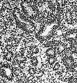
File:Medulloepithelioma_Histology.jpg | Medulloepithelioma/ETMR ([[H&E]]) | |||
File:Ependymoblastoma_ETMRjpg.jpg | Medulloepithelioma/ETMR ([[H&E]]) | |||
File:Medulloepitheliom_high.jpg | Medulloepithelioma/ETMR ([[H&E]]) | |||
</gallery> | |||
===Ependymoblastoma=== | |||
*Often supratentorial, well circumscribed. | |||
*Multilayered ("ependymoblastous") rosettes. | |||
*High mitotic and proliferative activity | |||
*Ependymoblastoma are grouped with medulloepithelioma and [[ETANTR]] into embryonal tumors with multilayered rosettes ([[ETMR]]).<ref name="pmid26438544">{{Cite journal | last1 = Horwitz | first1 = M. | last2 = Dufour | first2 = C. | last3 = Leblond | first3 = P. | last4 = Bourdeaut | first4 = F. | last5 = Faure-Conter | first5 = C. | last6 = Bertozzi | first6 = AI. | last7 = Delisle | first7 = MB. | last8 = Palenzuela | first8 = G. | last9 = Jouvet | first9 = A. | title = Embryonal tumors with multilayered rosettes in children: the SFCE experience. | journal = Childs Nerv Syst | volume = | issue = | pages = | month = Oct | year = 2015 | doi = 10.1007/s00381-015-2920-2 | PMID = 26438544 }}</ref> | |||
<gallery> | |||
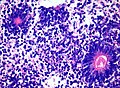
File:Ependymoblastoma.jpg | Ependymoblastoma. (WC/AFIP) | |||
File:Ependymoblastomatous_Rosette.jpg | Ependymoblastous rosettes. | |||
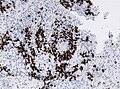
File:MIB1_ependymoblastoma.jpg | MIB-1 in ependymoblastous rosettes. | |||
</gallery> | |||
==Immunohistochemistry== | |||
* S-100 +ve. | |||
* [[INI1]] +ve (loss defines tumour as [[ATRT]]). | |||
* [[LIN28]]+ve (in [[ETMR]]), otherwise -ve. <ref>{{Cite journal | last1 = Korshunov | first1 = A. | last2 = Ryzhova | first2 = M. | last3 = Jones | first3 = DT. | last4 = Northcott | first4 = PA. | last5 = van Sluis | first5 = P. | last6 = Volckmann | first6 = R. | last7 = Koster | first7 = J. | last8 = Versteeg | first8 = R. | last9 = Cowdrey | first9 = C. | title = LIN28A immunoreactivity is a potent diagnostic marker of embryonal tumor with multilayered rosettes (ETMR). | journal = Acta Neuropathol | volume = 124 | issue = 6 | pages = 875-81 | month = Dec | year = 2012 | doi = 10.1007/s00401-012-1068-3 | PMID = 23161096 }}</ref> | |||
* Nestin +ve | |||
* [[MAP2]] +ve/-ve | |||
* Vimentin +ve | |||
* [[IDH-1]] -ve | |||
* No [[ATRX]] loss | |||
* MIB-1 between 20-80% (usu. 50%) | |||
==Molecular genetics== | |||
Divergent molecular subgroups are emerging: | |||
* Loss of 9q / CDKN2A deletions in CNS neuroblastoma<ref>{{Cite journal | last1 = Pfister | first1 = S. | last2 = Remke | first2 = M. | last3 = Toedt | first3 = G. | last4 = Werft | first4 = W. | last5 = Benner | first5 = A. | last6 = Mendrzyk | first6 = F. | last7 = Wittmann | first7 = A. | last8 = Devens | first8 = F. | last9 = von Hoff | first9 = K. | title = Supratentorial primitive neuroectodermal tumors of the central nervous system frequently harbor deletions of the CDKN2A locus and other genomic aberrations distinct from medulloblastomas. | journal = Genes Chromosomes Cancer | volume = 46 | issue = 9 | pages = 839-51 | month = Sep | year = 2007 | doi = 10.1002/gcc.20471 | PMID = 17592618 }}</ref> | |||
*Amplification 19q13.42 in [[ETMR]]<ref>{{Cite journal | last1 = Korshunov | first1 = A. | last2 = Remke | first2 = M. | last3 = Gessi | first3 = M. | last4 = Ryzhova | first4 = M. | last5 = Hielscher | first5 = T. | last6 = Witt | first6 = H. | last7 = Tobias | first7 = V. | last8 = Buccoliero | first8 = AM. | last9 = Sardi | first9 = I. | title = Focal genomic amplification at 19q13.42 comprises a powerful diagnostic marker for embryonal tumors with ependymoblastic rosettes. | journal = Acta Neuropathol | volume = 120 | issue = 2 | pages = 253-60 | month = Aug | year = 2010 | doi = 10.1007/s00401-010-0688-8 | PMID = 20407781 }}</ref> | |||
DDx: | |||
* Small round blue cell tumours | |||
* [[Medulloblastoma]] | |||
* [[ATRT]] (INI1 loss) | |||
* Anaplastic [[ependymoma]] (RELA fusions) | |||
* Paediatric [[glioblastoma]] (IDH1/2) and (H3F3A mutations) | |||
*[[Embryonal tumour with abundant neuropil and true rosettes]] (ETANTR) - currently no distinct WHO entity.<ref name=pmid19563506>{{cite journal |author=Buccoliero AM, Castiglione F, Degl'Innocenti DR, ''et al.'' |title=Embryonal tumor with abundant neuropil and true rosettes: morphological, immunohistochemical, ultrastructural and molecular study of a case showing features of medulloepithelioma and areas of mesenchymal and epithelial differentiation |journal=Neuropathology |volume=30 |issue=1 |pages=84–91 |year=2010 |month=February |pmid=19563506 |doi=10.1111/j.1440-1789.2009.01040.x |url=}}</ref> | |||
===Images=== | |||
www: | |||
*[http://path.upmc.edu/cases/case414.html Primitive neuroectodermal tumour - several images (upmc.edu)]. | |||
*[http://path.upmc.edu/cases/case649.html GBM with PNET component - several images (upmc.edu)]. | |||
==See also== | |||
*[[Neuropathology tumours]]. | |||
==References== | |||
{{Reflist|2}} | |||
[[Category:Neuropathology tumours]] | |||
[[Category:Embryonal tumour]] | |||
[[Category:WHO Grade IV tumour]] | |||
Latest revision as of 13:44, 4 October 2017
| Primitive neuroectodermal tumour | |
|---|---|
| Diagnosis in short | |
 CNS primitive neuroectodermal tumour H&E stain. | |
|
| |
| Synonyms | CNS-PNET |
| LM DDx | small round blue cell tumours |
| IHC | S-100 +ve, Syn +/-ve |
| Site | brain, spinal cord |
|
| |
| Prevalence | rare - typically in young adults |
| Prognosis | poor (WHO Grade IV) |
CNS Primitive neuroectodermal tumour, abbreviated CNS-PNET, is an abandoned neuropathology tumour description within in the group of embryonal tumours.
The terminology was introduced in 1973 [1] and used in the WHO 2007 classification of CNS tumors. Since 2016 this category has been replaced by the designation other CNS embryonal tumors.
General
- Should not be confused with peripheral primitive neuroectodermal tumour (abbreviated pPNET[2]), AKA Ewing sarcoma.
- The former category contained a heterogenous group of poorly differentiated WHO grade IV tumours associated with following ICD-O codes:
- 9473/3 CNS-PNET, NOS.
- 9500/3 CNS neuroblastoma.
- 9490/3 CNS ganglioneuroblastoma.
- 9501/3 Medulloepithelioma.
- 9392/3 Ependymoblastoma.
- Mainly children and adolescents.
- Cerebral hemisphere, brain stem or spinal cord.
- Cerebrospinal dissemination found in up to 1/3 patients.[3]
- Very poor prognosis[4]
Microscopic
Features:
- Small round blue cell tumour.
- Focal differentation into astrocytic, neuronal or ependymal phenotypes possible.
- May have true rosettes (slit-like/oval).
- Growth in streams or palisades possible ("spongioneuroblastoma").
- Vascular endothelial proliferations.
- Fibrillary background in tumours with advanced neuronal maturation (ganglioneuroblastomas).
- Variable mitotic activity.
Supratentorial PNET
- This category of small round- and blue cell tumor was used in the WHO 2007 CNS tumor classification to separate them from medulloblastomas.
- Tumors are today classified as AT/RT, Pineoblastoma, ETMR, H3F3A-mutated glioblastoma or CNS embryonal tumor, NOS.
CNS neuroblastoma
- Since WHO 2016 CNS classification this is now a subgroup of Other CNS embryonal tumours.
- Many CNS neuroblastoma / CNS ganglioneuroblastoma cluster molecularly into a group designated as CNS NB-FOXR2.[5]
- Usu Olig-2 +ve.
- Synaptophysin +ve.
CNS ganglioneuroblastoma
- This is now a subgroup of Other CNS embryonal tumours.
Lipomatous medulloblastoma
- These tumors are now designated as Cerebellar liponeurocytoma.
Melanotic medulloblastoma
- In WHO CNS 1997 still a distinct tumor.
- These tumors are now considered a variant of medulloblastoma.
Medullomyoblastoma
- Embryonal tumor with primitive neuronal cells and striated muscle component.
- First description in 1933.[6]
- Since WHO 2007 CNS tumor classification tumors were classified as medulloblastoma with myogenic differentiation.
Medulloepithelioma
- Neuroepithelial tumor cells arranged papillary, tubular or trabecular.
- Pseudostratified with PAS-positive membrane.
- Medulloepithelioma are grouped with ependymoblastomas and ETANTR into embryonal tumors with multilayered rosettes (ETMR).[7]
- Not the same tumour as the intraocular medulloepithelioma.[8]


Ependymoblastoma
- Often supratentorial, well circumscribed.
- Multilayered ("ependymoblastous") rosettes.
- High mitotic and proliferative activity
- Ependymoblastoma are grouped with medulloepithelioma and ETANTR into embryonal tumors with multilayered rosettes (ETMR).[7]
Ependymoblastoma. (WC/AFIP)
Ependymoblastous rosettes.
MIB-1 in ependymoblastous rosettes.


Immunohistochemistry
- S-100 +ve.
- INI1 +ve (loss defines tumour as ATRT).
- LIN28+ve (in ETMR), otherwise -ve. [9]
- Nestin +ve
- MAP2 +ve/-ve
- Vimentin +ve
- IDH-1 -ve
- No ATRX loss
- MIB-1 between 20-80% (usu. 50%)
Molecular genetics
Divergent molecular subgroups are emerging:
DDx:
- Small round blue cell tumours
- Medulloblastoma
- ATRT (INI1 loss)
- Anaplastic ependymoma (RELA fusions)
- Paediatric glioblastoma (IDH1/2) and (H3F3A mutations)
- Embryonal tumour with abundant neuropil and true rosettes (ETANTR) - currently no distinct WHO entity.[12]
Images
www:
- Primitive neuroectodermal tumour - several images (upmc.edu).
- GBM with PNET component - several images (upmc.edu).
See also
References
- ↑ Hart, MN.; Earle, KM. (Oct 1973). "Primitive neuroectodermal tumors of the brain in children.". Cancer 32 (4): 890-7. PMID 4751919.
- ↑ PST. 14 February 2011.
- ↑ Horten, BC.; Rubinstein, LJ. (Dec 1976). "Primary cerebral neuroblastoma. A clinicopathological study of 35 cases.". Brain 99 (4): 735-56. PMID 1030655.
- ↑ Tulla, M.; Berthold, F.; Graf, N.; Rutkowski, S.; von Schweinitz, D.; Spix, C.; Kaatsch, P. (Sep 2015). "Incidence, Trends, and Survival of Children With Embryonal Tumors.". Pediatrics 136 (3): e623-32. doi:10.1542/peds.2015-0224. PMID 26304823.
- ↑ Sturm, D.; Orr, BA.; Toprak, UH.; Hovestadt, V.; Jones, DTW.; Capper, D.; Sill, M.; Buchhalter, I. et al. (Feb 2016). "New Brain Tumor Entities Emerge from Molecular Classification of CNS-PNETs.". Cell 164 (5): 1060-1072. doi:10.1016/j.cell.2016.01.015. PMID 26919435.
- ↑ Brody, BS.; German, WJ. (Oct 1933). "Medulloblastoma of the Cerebellum: A Report of 15 Cases.". Yale J Biol Med 6 (1): 19-29. PMID 21433586.
- ↑ 7.0 7.1 Horwitz, M.; Dufour, C.; Leblond, P.; Bourdeaut, F.; Faure-Conter, C.; Bertozzi, AI.; Delisle, MB.; Palenzuela, G. et al. (Oct 2015). "Embryonal tumors with multilayered rosettes in children: the SFCE experience.". Childs Nerv Syst. doi:10.1007/s00381-015-2920-2. PMID 26438544.
- ↑ Korshunov, A.; Jakobiec, FA.; Eberhart, CG.; Hovestadt, V.; Capper, D.; Jones, DT.; Sturm, D.; Stagner, AM. et al. (Jul 2015). "Comparative integrated molecular analysis of intraocular medulloepitheliomas and central nervous system embryonal tumors with multilayered rosettes confirms that they are distinct nosologic entities.". Neuropathology. doi:10.1111/neup.12227. PMID 26183384.
- ↑ Korshunov, A.; Ryzhova, M.; Jones, DT.; Northcott, PA.; van Sluis, P.; Volckmann, R.; Koster, J.; Versteeg, R. et al. (Dec 2012). "LIN28A immunoreactivity is a potent diagnostic marker of embryonal tumor with multilayered rosettes (ETMR).". Acta Neuropathol 124 (6): 875-81. doi:10.1007/s00401-012-1068-3. PMID 23161096.
- ↑ Pfister, S.; Remke, M.; Toedt, G.; Werft, W.; Benner, A.; Mendrzyk, F.; Wittmann, A.; Devens, F. et al. (Sep 2007). "Supratentorial primitive neuroectodermal tumors of the central nervous system frequently harbor deletions of the CDKN2A locus and other genomic aberrations distinct from medulloblastomas.". Genes Chromosomes Cancer 46 (9): 839-51. doi:10.1002/gcc.20471. PMID 17592618.
- ↑ Korshunov, A.; Remke, M.; Gessi, M.; Ryzhova, M.; Hielscher, T.; Witt, H.; Tobias, V.; Buccoliero, AM. et al. (Aug 2010). "Focal genomic amplification at 19q13.42 comprises a powerful diagnostic marker for embryonal tumors with ependymoblastic rosettes.". Acta Neuropathol 120 (2): 253-60. doi:10.1007/s00401-010-0688-8. PMID 20407781.
- ↑ Buccoliero AM, Castiglione F, Degl'Innocenti DR, et al. (February 2010). "Embryonal tumor with abundant neuropil and true rosettes: morphological, immunohistochemical, ultrastructural and molecular study of a case showing features of medulloepithelioma and areas of mesenchymal and epithelial differentiation". Neuropathology 30 (1): 84–91. doi:10.1111/j.1440-1789.2009.01040.x. PMID 19563506.